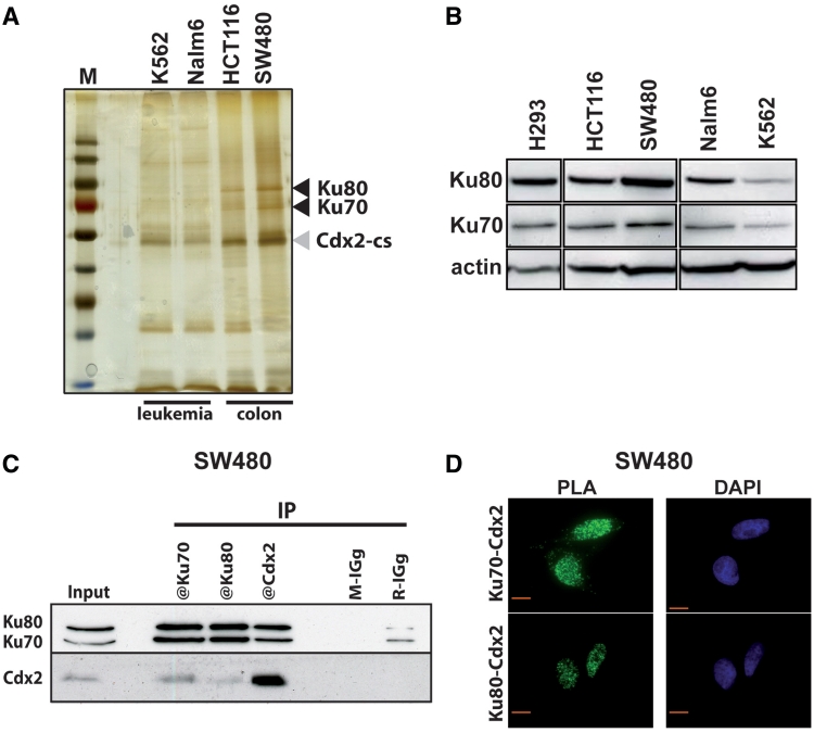
Figure 1.
Cdx2 interacts with Ku70/Ku80 proteins. (A) Silver-stained protein gel obtained after tandem affinity purification from K562, Nalm6, HCT116 and SW480 cells transfected with pCTAP-Cdx2. M: molecular weight protein ladder. Cdx2-CBP-SBP (Cdx2-cs) and Ku70/Ku80 proteins are indicated with gray or black arrowheads, respectively. (B) Western blot revealing the presence of Ku70 and Ku80 proteins in all studied cell lines. Nearly 20 µg of total proteins were separated on a gradient SDS–PAGE. Actin was used as loading control. (C) Coimmunoprecipitation of endogenous Cdx2 and Ku proteins. SW480 extracts were prepared and Cdx2, Ku70 or Ku80 were immunoprecipitated using corresponding antibodies. Mouse IGg (M-IGg) or rabbit IGg (R-IGg) were used as negative control. Western blot was revealed using rabbit anti-Ku70 and rabbit anti-Ku80 in the upper panel and using mouse anti-Cdx2 in the lower panel. About 20 µg of protein extracts were loaded in the input line. (D) In situ proximity ligation assay. Mouse anti-Cdx2 and rabbit anti-Ku70 (left panel) or mouse anti-Cdx2 and rabbit anti-Ku80 antibodies (right panel) were used to reveal endogenous proteins in SW480 cells. Green fluorescence corresponds to the PLA positive signal and indicates that the two molecules belong to the same protein complex; blue fluorescence corresponds to nuclei (DAPI staining). Red bar is 10 µm.

