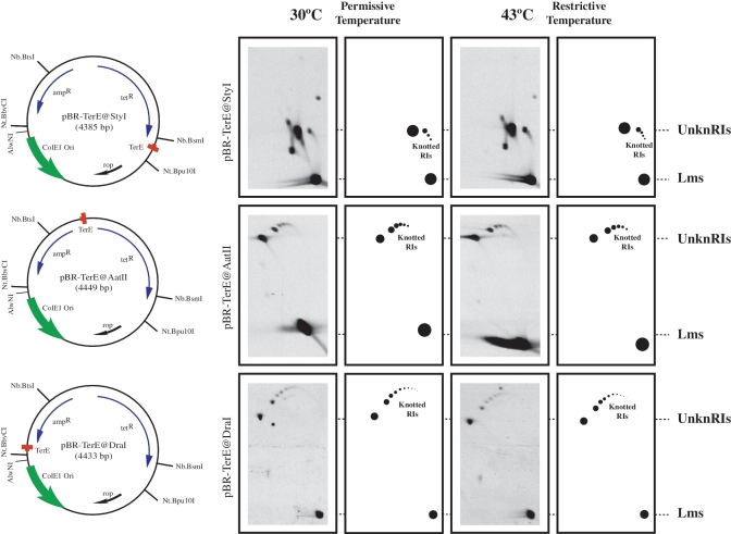
Figure 2.
Identification of partially replicated molecules at different stages of replication containing interchromatid knots. The name, mass and genetic maps of the plasmids used are indicated on the left side. Inside, each map shows the relative position of its most relevant features: the ColE1 unidirectional origin (ColE1 Ori), the E. coli terminator sequence (TerE) and the ampicillin- and tetracycline-resistance genes (ampR and tetR). Outside, the relative positions of sites recognized by specific restriction endonucleases are indicated. Autoradiograms of plasmid DNAs isolated from parE10 cells grown at the permissive and restrictive temperatures after digestion with AlwNI and analysed in 2D gels with their corresponding interpretation diagrams are shown on the right side. For comparison autoradiograms were aligned according to the electrophoretic mobility of unknotted replication intermediates (UnknRIs) and linearized molecules (Lms). Note that no significant difference was observed between the samples taken from cells grown at the permissive or restrictive temperatures.

