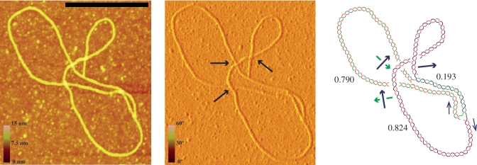
Figure 5.
Atomic force microscopy (AFM) allows the unambiguous identification of the shape of individual molecules extended on a flat surface. Visualization of height-data and phase-data (left and middle photographs, respectively) distinguishes which branch is above and which below at each individual node (see blue arrows in the middle photograph). This is essential to determine whether the molecule is knotted or not. The interpretative diagram shown to the right confirmed that this molecule corresponding to pBR-TerE@DraI was unknotted. In the diagram the parental duplex is drawn in blue and green whereas the nascent strands are drawn in red. Numbers indicate the relative size of each arm. Black arrows mark directionality of the sister duplexes and green and blue arrows indicate handedness. The scale on the left of the photographs represents height and phase and the black bar is 250-nm long.

