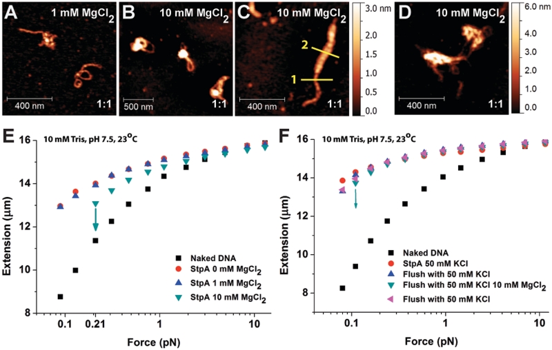
Figure 6.
Magnesium chloride (MgCl2) promotes DNA compaction via inter-co-filament interaction. (A–D) AFM images of 1:1 StpA:DNA complexes (300 nM StpA) incubated in buffer containing 1 or 10 mM MgCl2. The StpA/DNA ratios are at the bottom right corner of each sub panel: (A) In 1 mM MgCl2, linearized φX174 DNA substrates assumes similar rigid DNA hairpin structures to Figure 1A. (B and C) In 10 mM MgCl2, the DNA is organized into higher order aggregations. AFM width measurement showed the StpA-coated DNA has an apparent width of 20.44 nm (yellow line 1) while the thicker portion has an apparent width of 51.68 nm (yellow line 2). Considering the ∼12 nm AFM tip widening effect, the thicker portion is around four times as thick as the thinner portion, which is possibly caused by bundling of four DNA–StpA co-filaments. (D) In 10 mM MgCl2, linear 576-bp DNA substrate with the same StpA:DNA ratio and StpA concentration is organized into aggregates. (E) Force-extension curves of 600 nM StpA in varying MgCl2 buffer conditions from 0 to 10 mM MgCl2. StpA-induced DNA stiffening was observed at all non-zero MgCl2 concentrations. StpA-induced DNA folding events occurred at 10 mM MgCl2, as indicated by downward arrows. Before folding, DNA was stiffened indicated by the longer extension comparing with the naked DNA. (F) Force-extension curves of DNA fully coated with a StpA filament in the absence of surrounding free proteins in a series of buffer cycling. After the DNA folded at 10 mM MgCl2, it was completely unfolded at high force and then switched to 50 mM KCl, whereupon it was again completely stiffened with no folding. The DNA did not undergo any folding events in the absence of magnesium even when held at the lowest force (∼0.08 pN) up to 20 min (Supplementary Figure S6C). This result verifies that the observed DNA compaction by StpA was due to the presence of magnesium.

