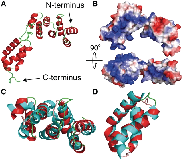
Figure 3.
Structure of Gln4(1–187) with comparisons to domains in S. aureus GatB (PDB ID: 3IP4). (A) Crystallographic structure of Gln4 residues 1–187 in cartoon representation. The proposed hinge region (Gly112Val113Gly114) is highlighted together with the likely interacting residue Trp160, and shown in stick representation. (B) Surface electrostatic model of Gln4 residues 1–187, shown with two orientations rotated by 90° relative to each other, with positively charged residues colored blue. (C and D) Structural alignment of helical and tail domains of Gln4 NTD and S. aureus GatB (PDB ID: 3IP4) (45). (C) The crystal structure of Gln4(1–110) (red) is superposed to the helical domain of GatB(295–406) (cyan). (D) The crystal structure of Gln4(119–178) (red) is superposed on the tail domain of GatB(414–475) (cyan).

