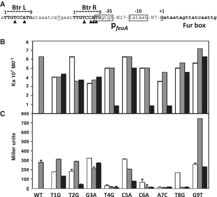
Figure 1.
Characterization of the Btr binding site. (A) Sequence of PfeuA showing the Btr binding site as a 9-bp direct repeat. A capital underlined T between the repeats was changed from G to create a restriction site (for generation of double mutants). The −35 and −10 elements are boxed and are separated by a 17-bp spacer region. The transcription start site is G (marked as +1). Closed triangles show positions critical for Btr:BB-mediated feuA activation. (B) Association constants (Ka) of Btr:BB binding to WT PfeuA (left most gray bar) and PfeuA mutants (single mutation in left repeat shown in white, single mutation in right repeat in gray, and the corresponding double mutation in black) as indicated underneath panel C. In each case, the binding affinity was measured by EMSA in the presence of 0.2 µM BB (see Supplementary Figure S1 for raw data). (C) Expression from PfeuA WT and mutants in vivo was monitored using a PfeuA-lacZ reporter integrated into strain HB8242 which lacks Fur and constitutively expresses BB (25). Mutations are indicated as in panel B (each set of three represents mutations in the left repeat, right repeat and both, respectively).

