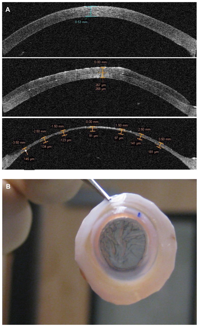
Figure 1.
Visante anterior segment optical coherence tomography image (A) showing initial cornea graft thickness (prior to epithelial removal) (top), after a 200 μm head pass (middle), and after the second 110 μm pass (bottom). After the double-passes, the cornea scleral rim shows striae as evidence of the thinness of less than 100 μm (B).

