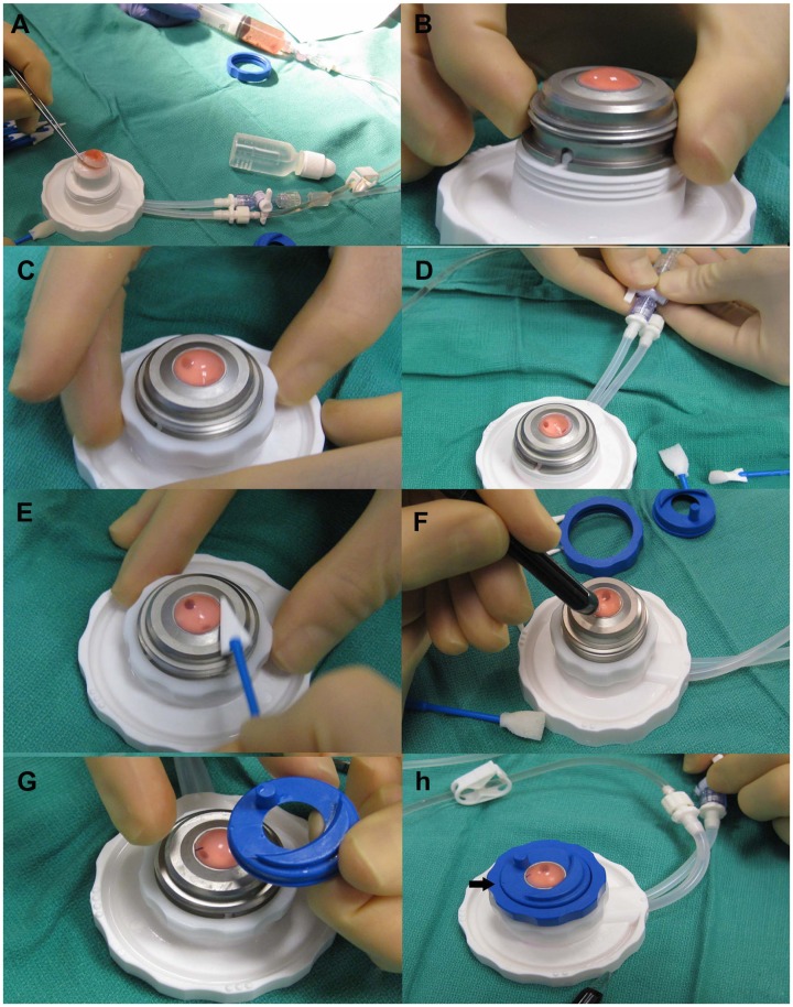
Figure 3.
(A) The cornea scleral button is centered on the anterior chamber while infusing Optisol through a syringe. (B) Once centration is achieved, the metal cover is placed over the cornea so that the groove fits into the white notch on the base of the anterior chamber. (C) A white locking ring is then tightened. (D) The balanced salt solution port is opened and then closed to equilibrate the pressure. (E) Epithelium is removed with a Weck-cell. (F) Pachymetry is measured in four quadrants to determine the thickest area of the cornea. (G) The guide ring is oriented so that the reference dot (arrow) is aligned with the alignment mark which is placed in the thickest quadrant of the cornea. (H) The stopcock to the balanced salt solution infusion is opened for a few seconds to equilibrate the pressure again prior to the first pass.

