
Figure 4.
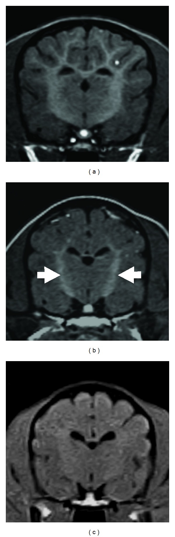
Transverse T1W images at the level of the thalamus/3rd ventricle. (a) An age-matched control dog at 2 months of age, (b) an affected laboratory dog at 2 months of age, (c) clinical case (animal 1) at 3 months of age. Symmetrical T1W hyperintensity was found in the internal capsule (arrows) in the affected laboratory dog (b), whereas there was no apparent hyperintensity in animal 1 (c). However, this internal capsule T1W intensity in the affected dog was not higher than that observed in the control dog (a).