
Figure 8.
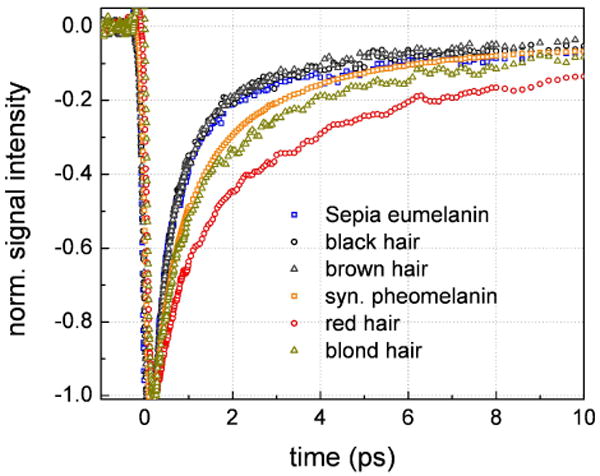
Baselined and normalized transient absorption decays for various non-human and human melanins. In this case the signals were normalized to have the same value at early time (200 fs) in order to compare the short-lived transients. The signals exhibit variability for different pheomelanins while they are virtually identical for different eumelanins.