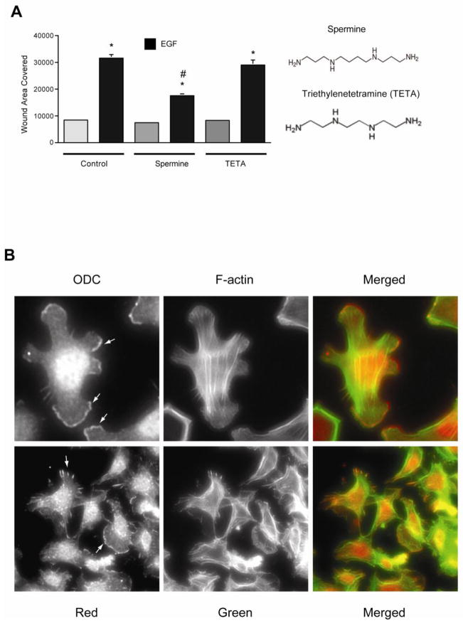
Fig. 2. Spermine inhibits EGF-induced migration.
A, confluent monolayers of IEC-6 cells were wounded with a gel loading tip in the center of the plates, washed and left untreated or treated with 10 nM EGF in the presence and absence of 10 μM of spermine or triethylenetetramine (TETA). Migration was calculated as described in the methods. Values are means ± SEMs of triplicates. * Significantly different compared to UT group. B, serum starved IEC-6 cells were plated in DMEM containing dFBS and were allowed to attach and spread. Cells were washed, fixed and processed for localization of ODC and f-actin as described in methods. A cropped image of a cell showing ODC localization in lamellipodia is shown (upper panel), A group of cells showing ODC and F-actin in lamellipodia and focal plaques (lower panel). Representative images from three experiments are shown.

