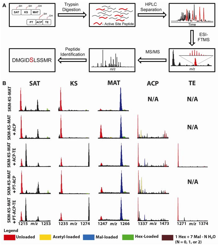
Figure 3.
Analysis of enzyme-bound PksA intermediates. A) Methods schematic. Selected proteins are combined in vitro with substrates. Following trypsin digest, peptides are separated by RP-HPLC in-line with ESI-FTMS to identify active site peptides and their acyl mass shifts. B) Summary of observed domain occupancies of SAT, KS, MAT, ACP, and TE active site peptides for hexanoyl- and malonyl-CoA reactions. Identities of the bound acyl-species are color-coded.

