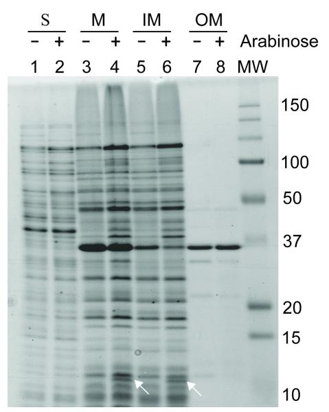
Figure 2. Localization of YgfX in the inner membrane.
E. coli BL21 (DE3) cells were transformed with pBAD24-ygfX (lane 2, 4, 6, and 8) or with pBAD24 (lane 1, 3, 5, and 7), and treated with 0.2% arabinose for 1 hr. The soluble (S) fractions (lane 1 and 2) and the membrane (M) fractions (lane 3 and 4) were separated by ultracentrifugation at 100,000 x g for 1 hr. The membrane fraction was resuspended in 1% N-laurylsarcosine (Hobb et al., 2009). The detergent-soluble inner membrane (IM; lane 5 and 6) and the insoluble outer membrane (OM; lane 7 and 8) were separated by ultracentrifugation as described above. Proteins were separated by 17.5% SDS-PAGE 447 and visualized by Coommassie blue staining.

