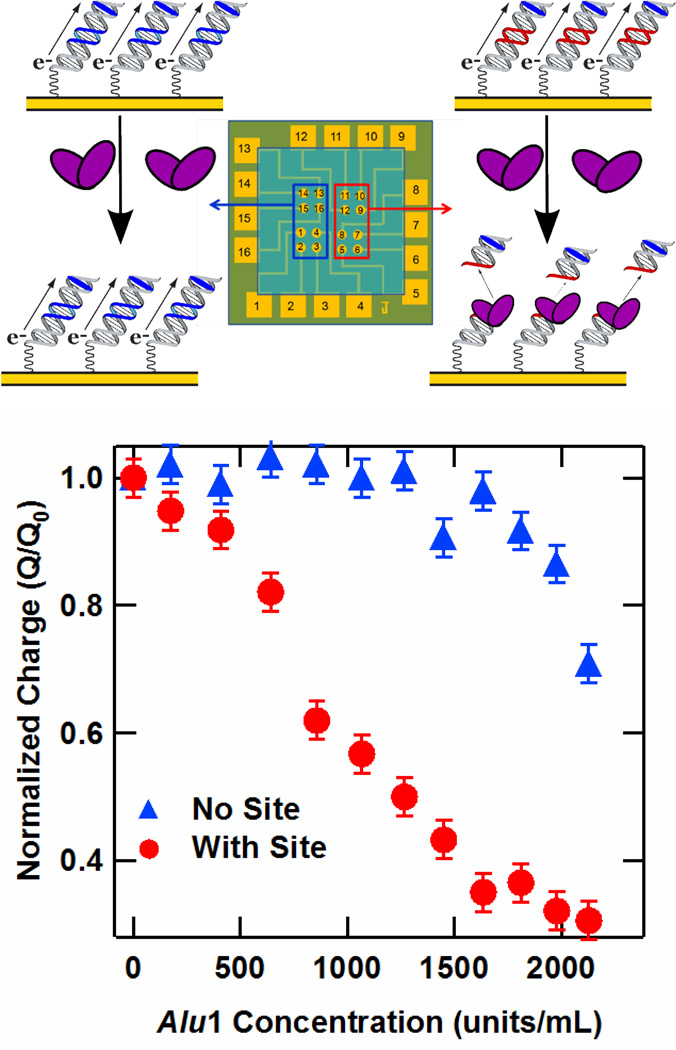
Figure 3.
Restriction assay on the DME chip. (Upper) Illustration of sequence-specific activity of the Alu1 restriction enzyme electrochemically monitored with a DME chip. (Lower) Charge versus Alu1 concentration for a DME chip for DNA with (Red) and without (Blue) the Alu1 restriction site. In particular, the sequence of the DNA containing the restriction site was the well matched 17-mer 5′-TNBGC GTC TCA GCT GAA GT-3′, where the italicized bases represent the restriction site and TNB is a thymine modified with a Nile Blue redox probe. The sequence absent this site but containing a three-base pseudosite was the well matched 17-mer 5′-TNBGC GTG CTT TAT ATC TC-3′, with the pseudosite given in italics. Charge was obtained by integrating the cathodic Nile Blue CV peaks obtained at a 50 mV/s scan rate after equilibration of the Alu1 activity at each concentration.

