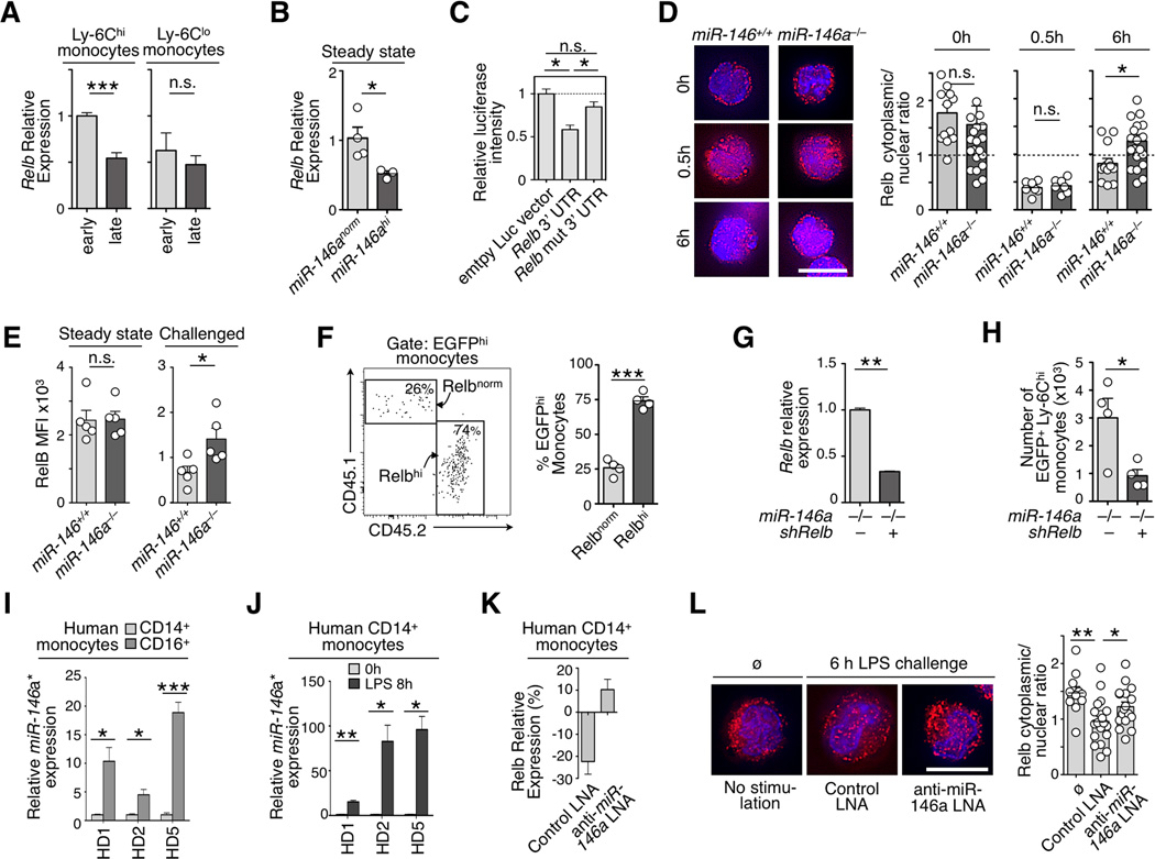
Figure 4. Relb is a miR-146a target in monocytes.
a) Relative Relb mRNA expression in monocytes subsets recruited to the peritoneal cavity at 2 h (early) and 8 h (late) post inflammatory challenge. Data are normalized to Ly-6Chi monocytes at 2 h (n=3).
b) Relative Relb mRNA expression in steady-state Ly-6Chi monocytes that over-express miR-146a (miR-146ahi) or not (miR-146anorm) (n=3).
c) Luciferase reporter assay for miR-146a–dependent regulation of Relb 3’ UTR. Luciferase activity was measured in NIH3T3 cells transfected with control empty vector, Relb 3’ UTR or a mutated version of the Relb 3’ UTR.
d) Immunofluorescence staining of Relb protein in wild-type or miR-146a−/− Ly-6Chi monocytes at 0, 0.5 and 6 h after LPS challenge. (Images are representative of n=7–21 cells analyzed per condition). Scale bar 10µm. Quantification shows cytoplasmic vs. nuclear fluorescence signal ratios.
e) Flow cytometry evaluation of intracellular Relb protein expression levels in wild-type or miR-146a−/− Ly-6Chi blood monocytes in steady-state or 6 h after LPS challenge (n=5).
f) Tracking of EGFP+ monocytes reconstituted with a Relb–overexpressing (Relbhi) or control (Relbnorm) vector in the peritoneal cavity 7 d after LPS challenge. Gating shows a representative result of the two competing monocyte populations (n=4 animals per group).
g) shRNA–mediated knockdown in EGFP+ cells measured by real-time PCR in miR-146a−/− Ly-6Chi monocytes (n=3).
h) Accumulation in the peritoneal cavity of EGFP+ miR-146a−/− monocytes transfected either with a shRelb or control construct 7 d after transfer into LPS challenged recipients.
i) Differential miR-146a* expression in CD14+ (CD16−) and CD16+(CD14−) monocytes from 3 healthy donors (HD) ex vivo.
j) Induction of miR-146a* in CD14+(CD16−) monocytes 6 h post LPS challenge (same donors as in i; n=3 technical replicates per group).
k) Percent change of Relb mRNA expression in CD14+(CD16−) monocytes of HD5 6 h post LPS challenge in presence of a scrambled or anti-miR-146a LNA (n=3).
l) Immunofluorescence staining of Relb protein in CD14+(CD16−) monocytes analyzed ex vivo (ø) or treated as in k. (Images are representative of n=11–19 cells analyzed per condition). Scale bar 10µm.
Quantification shows cytoplasmic vs. nuclear fluorescence signal ratios.
Data are presented as mean±SEM. (* p<0.05, ** p<0.001, *** p<0.0001, Student’s t-test).

