Figure 3.
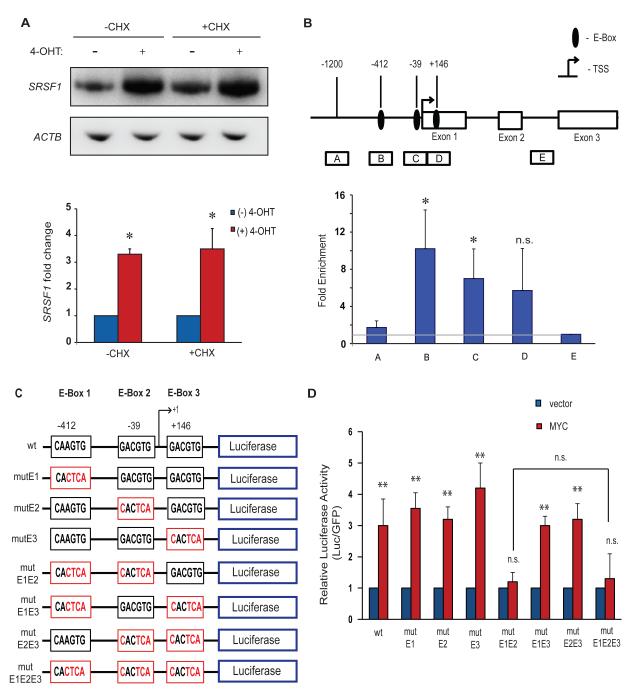
MYC binds to and activates the human SRSF1 promoter. (A) RT-PCR of IMR90-ER.MYC cells treated with 4-OHT, with or without cycloheximide. Error bars, s.d.; n= 3; **P<0.01. (B) MYC chromatin immunoprecipitation analysis at the SRSF1 promoter locus in the lung-carcinoma NCI-H460 cell line. Diagram of the SRSF1 gene indicating the E-boxes and amplicons (A-E) used for ChIP assays. The results are expressed as DNA enrichment in fragmented chromatin immunoprecipitated with anti-MYC antibody (relative to anti-rabbit IgG immunoprecipitation) and normalized to the amplicon E signal, as measured by quantitative PCR. The horizontal gray line represents no change in MYC-specific enrichment. Error bars, s.d.; n=3; t-test *P<0.05; n.s., not significant. (C) Diagram of the wild-type SRSF1 promoter, comprising three non-canonical E-boxes, and the E-Box mutants generated for reporter assays. Mutant E-boxes and residues are indicated in red. (D) Luciferase assay of reporter constructs in (C) co-transfected with MYC cDNA or vector control into NIH3T3 cells. Luciferase activity was normalized to co-transfected GFP, and the relative activity is plotted. Error bars, s.d.; n=3; t-test **P<0.01; n.s., not significant.

