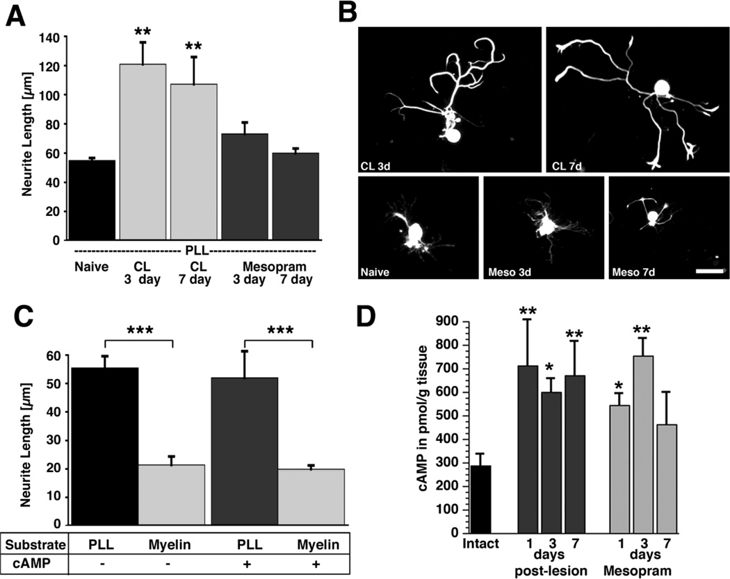
Figure 1. Quantification of in vitro neurite growth and cAMP levels in adult DRG neurons after conditioning lesions and infusion of phosphodiesterase inhibitors.
(A) Adult lumbar DRGs (L4–6) were dissected from naïve animals, animals that underwent a sciatic nerve crush 3 or 7 prior to isolation, or animals that received subcutaneous infusions of mesopram for 3 or 7 days prior to isolation. Quantification of neurite length indicates a significant increase in neurite outgrowth on poly-L-lysine (PLL) 3 and 7 days after conditioning lesions (**p<0.01 compared to naive), whereas infusions of the PDE IV inhibitor mesopram have no effect. (B) Examples of NF-200 labeled neurons indicate enhanced growth 3 days (CL 3d) and 7 days (CL 7d) after conditioning lesions but not after mesopram infusion (Meso 3d; Meso 7d). (C) db-cAMP (2 mM) does not increase neurite outgrowth of adult neurons on PLL or myelin. Myelin is strongly inhibitory in the presence and absence of db-cAMP (*** p<0.001 comparing PLL to myelin). Cells were cultivated for 72h and labeled for NF-200 to identify large and medium sized neurons. Data are presented as the means ± SEM of the average neurite length obtained in at least 3 independent experiments. (D) Quantification of cAMP levels in DRGs by ELISA. L4–6 DRGs were dissected from naïve animals, animals that underwent sciatic nerve crush lesions, and animals that received subcutaneous infusions of mesopram for the time indicated. Sciatic nerve lesions result in significant increases in cAMP levels at 1, 3 and 7 days post-lesion. Infusions of mesopram only lead to transient increases in cAMP levels (ANOVA followed by Fischer’s posthoc testing **p<0.01, * p<0.05, compared to naïve controls). Scale bar= 79 µm in (B).

