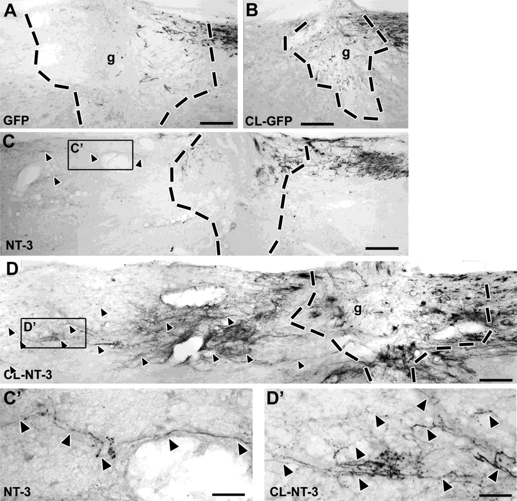
Figure 2. A combination of pre-conditioning lesions and lentiviral NT-3 gene transfer results in significant axonal bridging across the lesion site.
(A) CTB-labeled ascending sensory axons fail to bridge across a lesion site filled with BMSC in control animals that received injections of Lenti-GFP. (B) Conditioning lesions combined with control Lenti-GFP injections do not increase the number of bridging axons. (C) Animals that received Lenti-NT-3 rostral to a lesion (without conditioning lesions) show some bridging axons extending only for short distances (quantified in Fig. 4). (D) In contrast, a combination of Lenti-NT-3 and pre-conditioning lesions results in numerous axon bridging beyond the lesion and extension for longer distances. (C’, D‘) Higher magnification of boxed areas in (C) and (D), respectively, showing regenerating CTB-labeled axons (arrowheads) rostral to the lesion site. Rostral is to the left, dorsal to the top. Scale bar= 170 µm in (A–D), 42 µm in (C’, D’). Dashed lines indicate lesion/graft site determined by the absence of GFAP immunolabeling (see Fig. 3).

