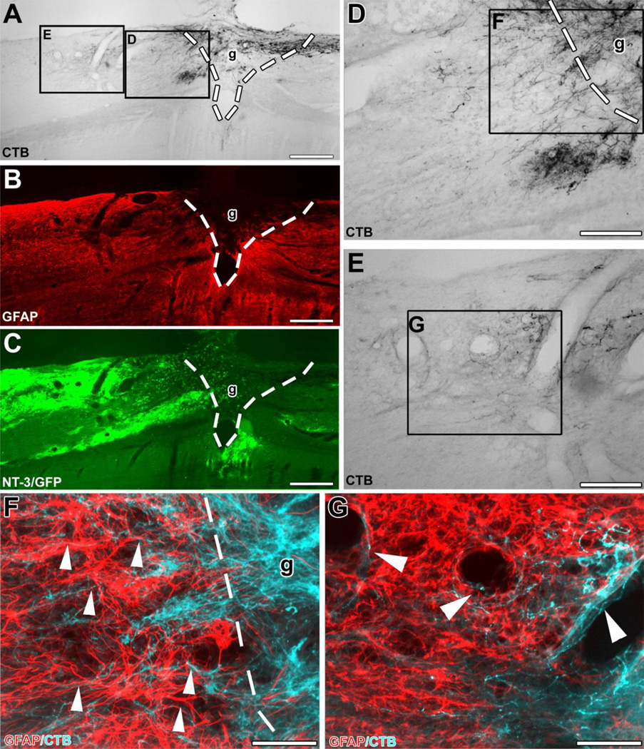
Figure 3. Ascending sensory axons extend beyond the graft/lesion site towards Lenti-NT-3-transduced cells in an animal that received pre-conditioning lesions and Lenti-NT-3 gene transfer.
Triple immunolabeling for (A) CTB to label ascending sensory axons, (B) GFAP to indicate the extent of the lesion/graft, and (C) GFP to label Lenti-NT-3-transduced cells in sagittal spinal cord sections. (D, E) Higher magnification of insets in (A) shows (D) axons growing across the rostral graft (g)/lesion site border (indicated by dashed lines). (E) Numerous axons are present further rostral to the lesion site, shown at higher magnification in (G). (F, G) Double immunolabeling for CTB-labeled axons (pseudocolored blue) and GFAP (red) at the (F) rostral host/graft interface (indicated by dashed lines) and (G) in the host spinal cord beyond the lesion. Bridging axons were often found to (F) orient along GFAP-labeled processes (arrowheads) and (G) were occasionally associated with blood vessels beyond the lesion site. Rostral is to the left, dorsal to the top. Scale bars 424 µm in (A–C), 170 µm in (D, E), 85 µm in (F, G).

