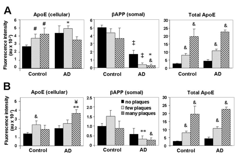
Figure 3.
βAPP and ApoE are differentially modulated in relation to plaque pathology. Intensities of βAPP fluorescence in neuronal somata, ApoE fluorescence in any cellular compartment, and total ApoE fluorescence were determined using analyses described in Materials and Methods. Fields were grouped according to “no plaques” (0), “few plaques” (1–4), or “many plaques” (≥5), in amygdala (A) or hippocampus (B). Values reflect the mean ± SEM from 13 (no plaques), 12 (few plaques), or 7 (many plaques) individuals. & p < 0.02, # p < 0.05 vs. control, no plaques; ‡ p < 0.001, ** p < 0.01, * p < 0.05 vs. corresponding control value; ¥ p < 0.01 vs. AD, no plaques.

