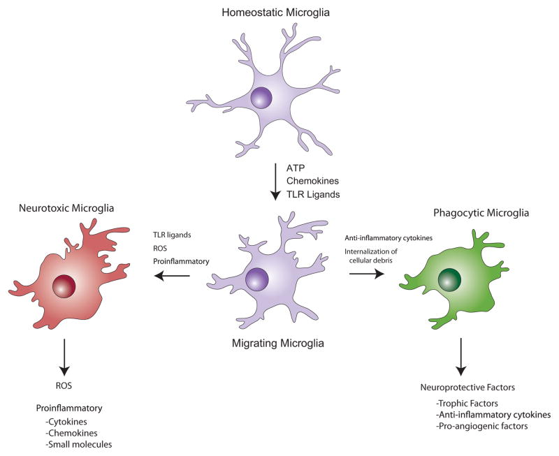
Figure 3. Microglia play divergent roles during neurodegeneration.
In the healthy CNS, microglia survey their immediate environment, and in this “resting state”, do not express inflammatory mediators. However, after exposure to a number of chemical signals from damaged neurons, microglia respond rapidly and physically migrate to the site of injury. Responding microglia may then adopt a pattern of behavior similar to pro-inflammatory macrophages (shown in red), as they release molecules intended to protect against pathogens, including neurotoxic cytokines, reactive oxygen species (ROS), and small molecules, such as quinolinic acid, that promote excitotoxicity. The release of cytokines and chemokines can lead to the recruitment of additional inflammatory cells from adjacent blood vessels, and may also engage astrocytes in the pro-inflammatory response. Alternatively, activated microglia may exhibit behaviors associated with anti-inflammatory macrophages (shown in green), secreting molecules that promote tissue repair, and internalizing cellular debris including aggregated, misfolded proteins such as β-amyloid, through phagocytosis. Whether two distinct populations of microglia exist that are committed to either of these response patterns, or all microglia can be induced to exhibit either response behavior when exposed to the correct combination of signals, remains to be determined.

