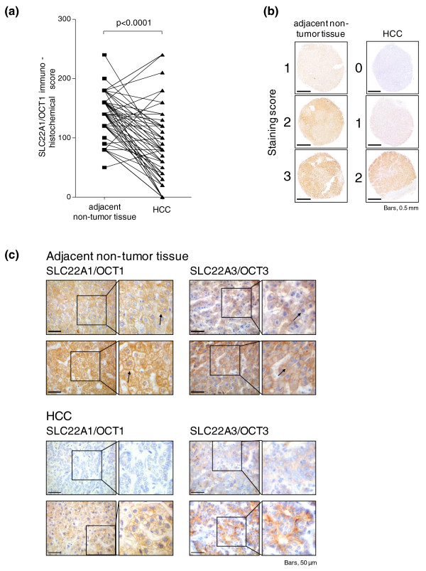
Figure 2.
Immunohistochemical staining of SLC22A1 protein in hepatocellular carcinoma and histological non-tumor adjacent tissue. (a) Expression of SLC22A1 is significantly decreased in hepatocellular carcinoma (HCC) versus adjacent non-tumor tissue (P < 0.0001) and individual paired results are given. For each patient, score values from both tissues are connected by a line. (b) Examples of different staining intensities (0 = negative, 1 = low, 2 = medium, 3 = high) in non-tumor as well as HCC tissues. SLC22A1 was present in all non-tumor tissues. (c) Exemplary weak immunohistochemical staining of SLC22A1 in two HCC samples is shown compared with strong staining in adjacent non-tumor liver tissue. In addition, representative examples of SLC22A3 staining in HCC tissue as well as in adjacent non-tumor liver tissue is shown. SLC22A1 and SLC22A3 are detected in the sinusoidal membrane of the hepatocytes (black arrows).

