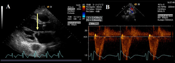
Figure 1.
Transthoracic echocardiogram - parasternal long-axis view with evidence of a nodular image attached to the atrial side of the anterior leaflet of the mitral valve (A, arrow) and a holodiastolic flow reversal in the descending thoracic aorta recorded from a suprasternal notch window with terminal velocity > 20 cm/s (B) supporting severe aortic regurgitation.

