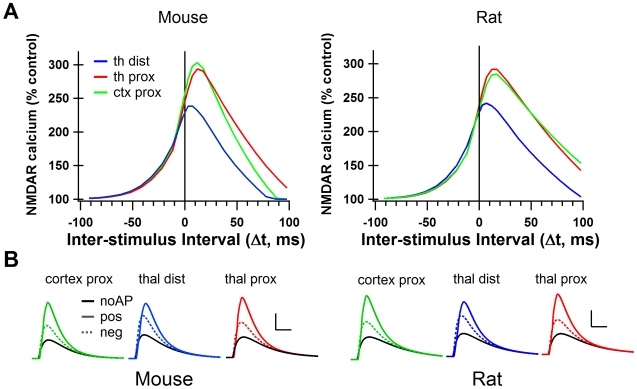
Figure 6. The cortico-striatal and thalamo-striatal synapses onto MSPNs have different synaptic characteristics.
A. Peak NMDAR calcium curves from simulations using cortico-striatal and thalamo-striatal parameters from mouse striatum (Ding et al., 2008) and from rat striatum (Smeal et al., 2008). Cortico-striatal curve for mouse is the same as the green trace in figure 2C. B. NMDAR-mediated calcium for positive (Δt = +11 ms, solid color), negative (Δt = −12 ms, dotted color) and no AP control conditions (solid black) under cortico-striatal and thalamo-striatal conditions in mouse and rat. Scale bars: 2 µM [Ca2+] vertical, 50 ms horizontal. thal dist = thalamo-striatal synapses stimulated on tertiary dendritic spines, thal prox = thalamo-striatal synapses stimulated on secondary dendritic spines. cortex prox = cortico-striatal synapses stimulated on secondary dendritic spines.

