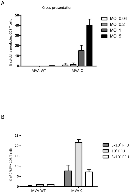
Figure 7. MVA-C infection induces antigen cross-presentation to CD8 T cells and T cell proliferation.
A) Human moDCs were incubated with apoptotic infected-HeLa cells before a CD8 T cell clone was added. After overnight incubation, cells were harvested and among the lymphocyte population, CD8 cells were gated and analyzed for IFN-γ, TNF-α, IL-2 and MIP-1β production. Cytokine production by HIV-specific CD8 T cells was determined. Percentages of CD8 T cells producing any cytokine are indicated at the various virus multiplicities. B) MVA-C and parental MVA-WT were evaluated in vitro using cryopreserved PBMCs from an HIV-1-infected subject. Cell proliferation using the CFSE dilution assay was measured 6 days after stimulation. At the end of the stimulation period, cells were stained for CD3, CD4, CD8 and a viability marker and analyzed by flow cytometry. Mean values and standard deviation of at least three experiments are shown in panels A and B.

