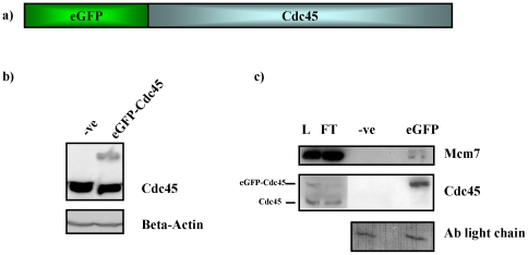
Figure 1. HeLa S3 cells stably expressing eGFP-Cdc45.
Panel a, schematic diagram of eGFP-Cdc45 protein encoded by Cdc45L ORF cloned into pIC113gw vector. Panel b, total cell extract HeLaS3 cells (-ve) and Hela S3 cells stably expressing eGFP-Cdc45 (eGFP-Cdc45) normalized for protein content and analysed by western blotting using antibodies raised against Cdc45 and β-Actin, which serves as a loading control. Panel c, western blot analysis of immunoprecipitation of eGFP-Cdc45 using GFP-Trap IP from HeLa S3 cells transiently expressing eGFP-Cdc45. Verification of purification of eGFP-Cdc45 and co-immunoprecipitation of Mcm7 was carried out using antibodies raised against Mcm7 and Cdc45. Input (L), unbound (FT), mock IP (-ve) and IP from cells expressing eGFP-Cdc45 (eGFP) indicate yield of the IP and co-immunoprecipiation. Antibody light chain acts as a loading control.

