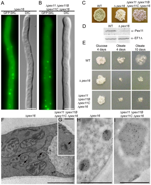
Figure 7. Pex16 is not essential for peroxisome biogenesis.
Fluorescence microscopy analysis of P. chrysogenum Δpex16 GFP.SKL (A) and Δpex11 Δpex11B Δpex11C Δpex16 GFP.SKL (B) cells. Cells were grown for 40 h in PPM. For corresponding WT see Fig. 3A. Scale bars represent 5 µm. C. Cells devoid of pex16 are sporulation deficient. Colonies of WT and both Δpex16 GFP.SKL and Δpex11 Δpex11B Δpex11C Δpex16 GFP.SKL strains were grown for 7 days on sporulation inducing R-agar plates. D. Δpex16 cells are characterized by decreased levels of Pex11. Western blots of WT and Δpex16 GFP.SKL cell extracts were prepared and decorated with α-Pex11 antibodies; translation elongation factor 1- (eEF1A) was used as a loading control. E. P. chrysogenum cells lacking pex16 are able to grow on oleic acid although at decreased rates. WT, Δpex16 GFP.SKL and Δpex11 Δpex11B Δpex11C Δpex16 GFP.SKL strains were grown for 10 days on mineral medium containing 0.5% glucose or 0.1% oleic acid as a sole carbon source. Electron micrographs of Δpex16 GFP.SKL (F) and Δpex11 Δpex11B Δpex11C Δpex16 GFP.SKL (G) cells grown for 40 h in PPM; P – peroxisome; M – mitochondrion; V – vacuole; arrows indicate protein dense inclusions. Scale bars represent 1 µm. Electron micrographs representing α-IAT immunolabelling of sections of Δpex16 GFP.SKL (H) and Δpex11 Δpex11B Δpex11C Δpex16 GFP.SKL (I) cells. Scale bars represent 1 µm.

