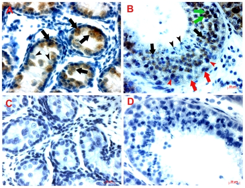
Figure 3. Immunohistochemical analysis of POU5F1 protein in prepubertal and adult testis sections.
(A) In 4-month-old testis section, most germ cells express POU5F1 (black arrows) and a few germ cells did not stain (arrowheads). POU5F1 expression is not seen in Sertoli and interstitial cells. (B) In 2-year-old testis section, POU5F1 expression is present in cytoplasm (black arrows) and nuclei of (green arrows) of round spermatids. At this age, spermatocytes (red arrowheads) and spermatogonia (red arrows) show very weak cytoplasmic localization of POU5F1 protein. Elongated spermatids show no expression of POU5F1 protein (black arrowheads). In a negative control (C, D), where primary antibody was omitted, no positive cells are present. Scale bar = 20 µm.

