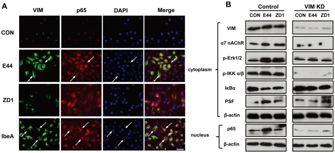
Figure 3. Role of vimentin in IbeA+ E. coli K1-induced NF-κB activation.
(A) Immunofluorescence microscopy was used to examine the correlation between vimentin reorganization and NF-κB translocation to the nucleus after 2 h of stimulation with IbeA protein (0.1 µg/ml), E44 or ZD1 (25 MOI). HBMECs were triple-stained with the V9 antibody against vimentin conjugated to FITC (green), the rabbit antibody against NF-κB (p65) conjugated to rhodamine (red), and DAPI (blue). The merged images are shown in the right-hand panels (Merge). Arrows indicated cells with colocalization of vimentin and NF-κB (p65) Scale bar, 50 µm. (B) Blockage of IbeA+ E. coli K1-induced NF-κB activation in HBMECs by siRNA-mediated knockdown of vimentin. HBMECs were transfected with vimentin or control siRNA as described in Materials and Methods. After 24 h incubation, the cells were treated with E44 or ZD1 (107/ml) for 30 min or 2 h. Vimentin (VIM), α7 nAChR, ERK1/2 phosphorylation (p-Erk1/2), IKK α/β phosphorylation (p-IKK α/β), IκBα degradation, and PSF re-localization were examined in cytoplasmic fractions after 30 min of stimulation with E. coli K1 strains. NF-κB (p65) translocation to the nucleus was examined in nuclear fractions after 2 h of incubation with E. coli K1 strains. β-actin in both fractions was detected as internal loading controls. Control: HBMECs transfected with control siRNA; VIM KD: HBMECs transfected with vimentin siRNA; UNT: Untreated HBMECs.

