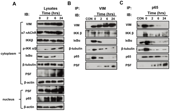
Figure 4. Time course analysis of IbeA-induced cytoplasmic activation and nuclear translocation of NF-κB.
HBMECs ware incubated with the IbeA protein (0.1 µg/ml) for 2, 6, and 24 h, respectively, and then the cytoplasmic and nuclear fractions were extracted. The cytoplasmic fractions were immunoprecipitated (IP) with the V9 anti-vimentin antibody and the rabbit anti-NF-κB (P65) antibody as described in Materials and Methods. The cytoplasmic and nuclear lysates (A), vimentin Co-IP complexes (B), and NF-κB (p65) Co-IP complexes (C) were subjected to western blot using the antibodies as described in Materials and Methods. CON: the IP control without primary antibodies incubation; 0 h: the control HBMECs without IbeA stimulation.

