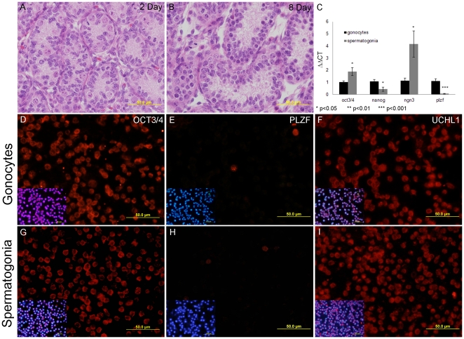
Figure 1. Characterisation of isolated gonocyte and spermatogonia cells.
Haematoxylin and eosin stained two day old testis section (1A) demonstrates that gonocytes are found in the centre of the seminiferous tubules of mice. Gonocytes differentiate and migrate to the basement membrane of the seminiferous tubules where they become established in their niche and begin the process of spermatogenesis as seen in the eight day old testis section stained with haematoxylin and eosin (1B). C) Total RNA from isolated germ cell populations (three biological replicates) reverse transcribed and subjected to qPCR. It was found that the gene expression of the germ cell markers Oct3/4 and Ngn3 were significantly higher (p<0.05) in spermatogonia, twice and four times the amount of gonocytes respectively. The gene expression of the ES cell markers Nanog and PLZF was significantly lower (p<0.05 and 0.001) in spermatogonia. Isolated gonocytes (D,E,F) and spermatogonia (G,H,I) were fixed on slides and stained with germ cell markers. 95% of both gonocytes (1D) and spermatogonia (1G) expressed oct4, while less than 5% of gonocytes (1E) and spermatogonia (1H) expressed PLZF. UCHL1 was expressed in over 95% of gonocytes (1F) and spermatogonia (1I). The expression of PLZF, OCT3/4 and UCHL1 are consistent with previous reports for germ cells indicating our isolated cell populations contained 95% germ cells.

