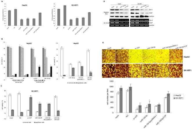
Figure 4. miR-129-5p could regulate the cell growth, apopotosis and migration of HCC cells dependent on the regulation of VCP expression.
A: HepG2 or SK-HEP1 cells were transfected and in the indicated time periods posttransfection, cell growth rate was evaluated using CCK8 assay. The result showed miR-129-5p and si-VCP could repress cell growth. The cell growth rate was restored after VCP expression vector were transfected into HepG2 or SK-HEP1 cells. Shown data are representative from three independent experiments. **:P<0.01. B: HepG2 cells were transfected and the apoptosis of cell was detected with FACS. Left panel showed miR-129-5p and si-VCP could increase the apoptosis of HepG2 cells at 48 h and 72 h after the transfection. Right panel showed the apoptosis of cell after miR-129-5p with/without VCP expression vector were transfected into HepG2 cells for 72 h. The experiment was repeated three times independently. NC: negative control RNA duplex; miR-129-5p: miR-129-5p mimic; miR-129-5p/pcDNA3.1: cells were co-transfected with miR-129-5p mimic and blank pcDNA3.1; miR-129-5p/VCP: cells were co-transfected with miR-129-5p mimic and VCP expression vector. The experiments were repeated three times independently.*:P<0.05. **:P<0.01. C: SK-HEP1 cells were transfected and the apoptosis of cells was detected with FACS. miR-129-5p and si-VCP could increase the apoptosis of SK-HEP1 cells at 72 h after the transfection. After the level of VCP was enhanced by transfecting VCP expression vector, the apoptosis rate of cells were returned. D:Detection the level of Bcl-2 and XIAP by RT-PCR and western blot. GAPDH served as the internal control. E:Transwell assay was performed to assess cell migration. Upper panel represented the photographs of treated and untreated cells at 24 h (×40 magnification). Lower panel showed the number of cells invaded at 24 h. Shown data are representative from four replicates per group. *P<0.05.

