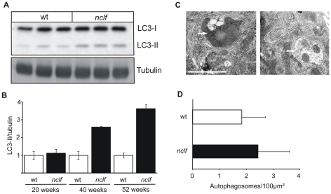
Figure 6. Accumulation of autophagosomes in nclf brain and hippocampal neurons.
A) Accumulation of autophagosomes was assessed by determining the levels of the autophagic marker protein microtubule-associated protein 1 light chain 3 -II (LC3-II) in wild-type or nclf total mouse brain extracts at 54 weeks of age by western blotting. B) Densitometric quantification of LC3-II levels normalized to tubulin as a loading control revealed enhanced autophagosome numbers with increasing age. Data are presented as mean ±SD, n = 3 per age. Wild-type values were set to 1. C) Double-membrane autophagic vacuoles (marked by arrows) were also found in hippocampal neurons from nclf mice cultured for 14 days. D) For quantification of autophagic vacuoles, pictures were taken from 37 randomly selected wild-type and nclf neurons of two different preparations. The number of autophagic vacuoles related to the cytoplasmic area was determined. Data are presented as mean ± S.E.M. Scale bars: 1 µm.

