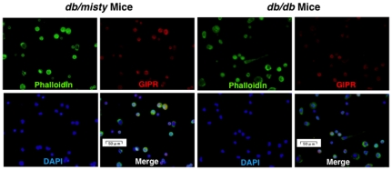
Figure 5. Expression of GIPR in exudate peritoneal macrophages from db/misty and db/db mice.
GIPR was stained with goat polyclonal anti-GIPR antibody followed by anti-goat Alexa Fluor 568. Phalloidin/DAPI staining shows F-actin cytoskeleton and nuclear morphology of mouse macrophages. These images were merged. Representative results are shown.

