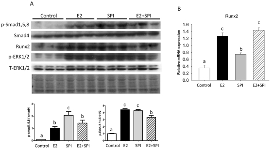
Figure 5. Cell signaling transduction pathway after activation of BMP-2 by SPI and E2 in bone.
(A), Protein was isolated from bone after aspiration of bone marrow cells and performed Western blots. Smad and ERK1/2 phosphorylation and Runx2 protein expression in pre-pubertal female rat bone from Control, standard casein diet group; E2, 10 µg/kg/d E2 treated group; SPI, soy protein isolate diet group; E2+SPI, combination of 10 µg/kg/d E2 treated and SPI diet group. Quantitation of the intensity of the phosphorylated bands of p-Smad1, 5, 8 and p-ERK1/2 in the autoradiograms were performed relative to expression of total Smad4 and ERK1/2. Blot of Ponceau S staining showing protein loading control for western blotting. (B), Showing real-time PCR analyses for Runx2. Data are Means ± S.E.M, n = 3, with different letters differ significantly from each other at p<0.05, a<b<c.

