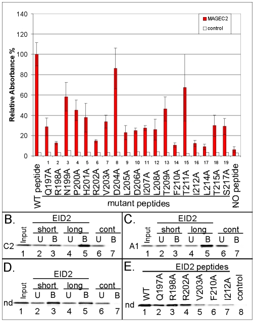
Figure 5. Analysis of EID2 binding to MAGE proteins.
(A.) Quantification of relative binding of the MAGEC2(129-339) protein (red columns) to the EID2 protein-based synthetic mutant peptides (listed in Table 1) using the PEPSCAN-ELISA method. Results show mean ± SEM of 3 independent measurements. His-hTRF2 protein (white column) was used in the control experiment. (B. to D.) The short (biotin-SGSG-201HRVDLDILTFTIALTAS217) and long (biotin-SGSG-197QRNPHRVDLDILTFTIALTAS217) EID2 peptides were pre-bound to the streptavidin-agarose beads and then incubated with in vitro translated MAGEC2 (aa 6-373; C2 in panel B.), MAGEA1 (aa 1-309; A1 in panel C.) and/or necdin (aa 1-321; nd in panel D.) protein, respectively. (E.) Wild type and selected EID2 mutant peptides (as indicated) were pre-bound to the streptavidin-agarose beads and then incubated with in vitro translated necdin protein. The reaction mixtures were analyzed by 15% SDS–PAGE gel electrophoresis. The amount of the in vitro translated proteins was measured by autoradiography. Control, no peptide.

