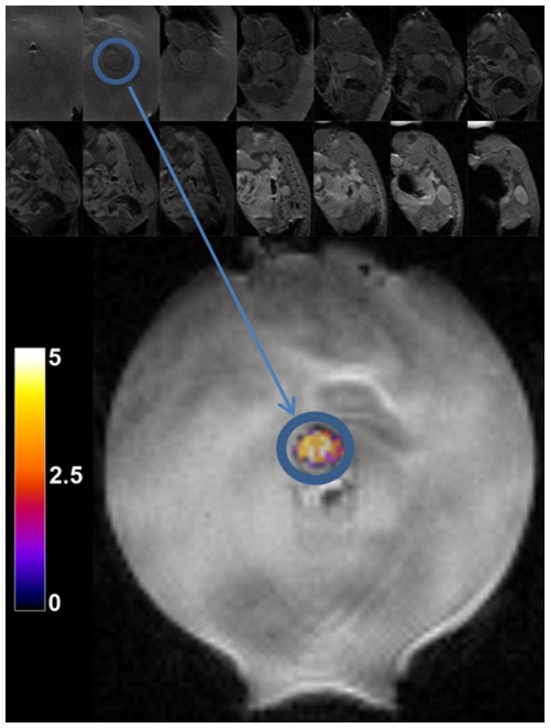
Figure 7. Localization and heating of Met-1 tumor.
Spin echo multi-slice MR magnitude images (acquired in the coronal plane) used to localize the Met-1 tumor prior to heating (top two rows). The animal is positioned on an acoustic gel pad. The gel pad has a central hole in which the tumor sits. Following localization of the tumor from the image series, the appropriate coronal plane is used for imaging and real-time temperature control. Temperature is overlaid in color on a magnitude MR image mid-way through the heating sequence (bottom).

