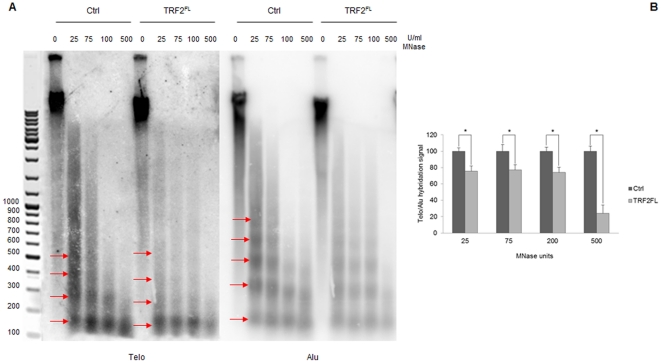
Figure 2. Altered nucleosome spacing at C33A telomeres.
(A) Digestion of chromatin from C33A cells infected with an empty vector and C33A cells overexpressing TRF2FL with increasing amounts of MNase. From the left: MNase digests separated on 1.5% agarose gel detected by hybridization with Telo probe; detection of telomeric nucleosomes after hybridization with Alu probe. (B) Ratio of the overall hybridization signal of the telomeric probe with respect to the Alu probe. Ratio values for control C33A cells have been normalized to 100. Error bars are s.d. of three independent experiments. Asterisks, p<0.05 based on unpaired Student's t-test.

