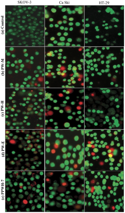
Figure 6. Morphological observation with AO/EB double staining by fluorescence microscope (200×).
Control cells and cells treated with PW-M, PW-H, PW-E and PPWH-7 at 10.0 µg/mL for 24 hours. Live cells with uniform bright green nuclei and cytoplasm were observed in control group (a). Apoptotic cells with condensed chromatin are uniformly fluorescent whereas fragmented nuclei and necrotic cells stained bright orange were observed in cells treated with the test agents studied (b–e). Images are representative of one of three similar experiments.

