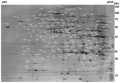
Figure 1. Representative 2-DE gel of Mycoplasma fermentans M64.
A 100 µg of total proteins were separated by a 24 cm pH 3–10 linear IPG strip for the first dimension IEF and a 24×20 cm 11–13% gradient gel for the second dimension SDS-PAGE. After silver staining, an average of 700 spots can be detected on a 2-DE gel. Spots are numbered according to the matched ORFs.

