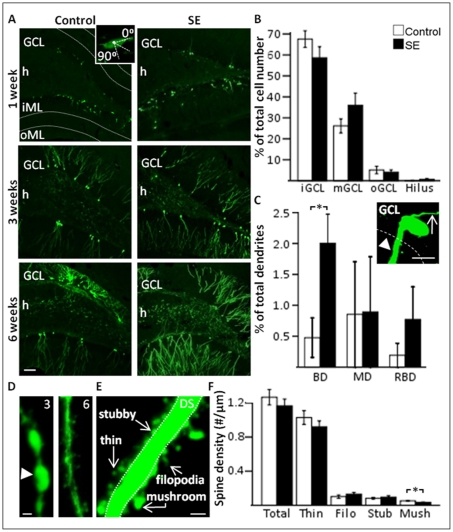
Figure 2. Neurons born in a partial SE (pSE)-induced environment develop with modest changes in gross morphology and dendritic spines.
A, Morphological and temporal development of GFP+ new cells formed after partial SE or in controls. Inset: horizontal polarisation compared to granule cell layer (GCL) of a GFP+ cell body at one week. B, Percentage of GFP+ cell bodies in inner, middle, and outer GCL (iGCL, mGCL, and oGCL, respectively), and the hilus at six weeks. C, Percentage of basal (BD), medial (MD) or recurrent basal dendrites (RBD) on GFP+ cells at six weeks, showing an increase in BD in pSE animals. Inset: GFP+ cell showing a BD projecting towards the hilus (arrow head) and an apical axon directed towards the ML (arrow). D, Photomicrograph showing representative dendritic beading at three weeks (arrow head) but not at six weeks. E, Representative image showing examples of the different types of dendritic spines in relation to the dendritic shaft (DS). F, Spine density on GFP+ dendrites from control and pSE animals, showing a decrease in number of mushroom spines. Means ± SEM, n = 7 for each group (morphological analysis), n = 8 control and n = 9 pSE group (dendritic spine analysis). *, P<0.05 unpaired t-test compared to control group. Scale bars are 50 µm (A), 10 µm (C), and 1 µm (D, E).

