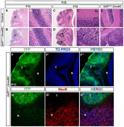
Figure 1. Shh pathway activation in Gdf7-lineage cells leads to cerebellar hyperplasia.
Gdf7Cre/+;SmoM2 gain-of-function mutant mice exhibit cerebellar defects. (A–B′) Hematoxylin-eosin staining of wild-type and Gdf7Cre/+;SmoM2 mutants. Prior to P10 histological sections of mutants are similar to control. (C–E′) Gdf7Cre/+;SmoM2 mutants over 14 days old develop ectopic foci of densely packed cells within the molecular layer of their cerebella. Higher magnification view of these foci reveals no discernible layer organization and resemblance to neoplastic lesions. Arrows in (D) indicate regions of hypercellularity. Boxed regions E and E″ are magnified and shown in the right adjacent panels. Arrows in D′ indicate nuclear molding. Arrowheads in D′ indicate apoptotic nuclei. (F-F″) The ectopic foci consist of cells of the Gdf7-lineage as indicated by their expression of SmoM2-YFP. (G-G″) Ectopic foci do not express differentiated neuronal marker NeuN. Abbreviations: EGL, external granular layer. ML, molecular layer. IGL, internal granular layer. T, tumor region. N, normal cerebellum.

