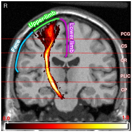
Figure 4. Segmented intracranial corticospinal tract (CST) atlas and the somatotopic arrangement within the primary motor cortex.
The probabilistic corticospinal tract (CST) atlas is overlaid onto the coronal orientation T1-MRI template (Montreal Neurological Institute). The color bar indicates the full range of the probability values in the CST atlas. The horizontal lines indicate the approximate anatomical levels at which the five segments were made along the length of the CST. PCG: Precentral gyrus, CS: Centrum semiovale, CR: Corona radiata, PLIC: Posterior limb of the internal capsule, CP: Cerebral peduncle and R: Right side of the subject. The blue, green and purple lines indicate the somatotopic arrangement within the primary motor cortex that are associated with the face and lower and upper limb parts, respectively.

