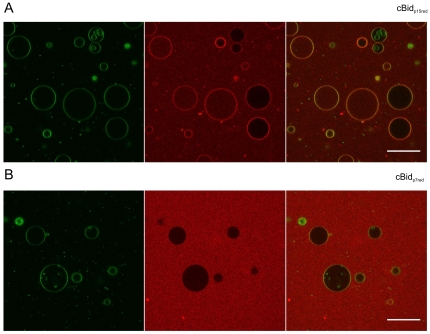
Figure 6. Binding of p15 and p7 to GUVs.
cBidp15red (C30S) and cBidp7red (126S) are shown in (A) and (B), respectively. GUVs composed of 80% phosphatidylcholine (egg), 20% cardiolipin (bovine heart) and <0.05%. DiO (Invitrogen) was incubated with 25 nM of the Alexa 633 labeled cBid variant. DiO is shown in the first panel (green) and the Alexa 633 labeled cBid variant in the second (red). The merge of the red and the green channel is shown in the third panel. The bar indicates 50 µm. Pictures were taken after 1 h. Due to better visualization the brightness in the red channel was increase for cBidp7red (126S). Notably, due to the fragility of the GUVs, 10–20% of the total GUVs are permeabilized even in absence of any protein.

