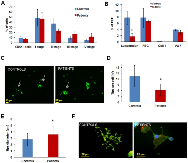
Figure 1. Megakaryocyte differentiation and proplatelet formation.
(A) Megakaryocyte maturation stages of patients did not differ from those of controls. (B) Proplatelet formation (PPF) from patient megakaryocytes in suspension was drastically reduced. When megakaryocytes were plated on type I collagen proplatelet formation was absent, similar to controls, while on fibrinogen and von Willebrand factor the number of megakaryocytes extending proplatelets was normal. *p<0.05 vs control. (C) Representative pictures of proplatelet formation in suspension, in a control subject and a patient (20× magnification). Arrows indicate pro-platelets, only one developing proplatelet is evident in the patient sample. (D, E) Patient megakaryocytes extended a reduced number of proplatelets with abnormal characteristics: a spread shape with shorter than normal proplatelet shafts and tips significantly decreased in number and larger in size than those of controls. *p<0.05 vs control. (F) Representative images of megakaryocytes from patients and controls releasing proplatelets upon adhesion to fibrinogen.

