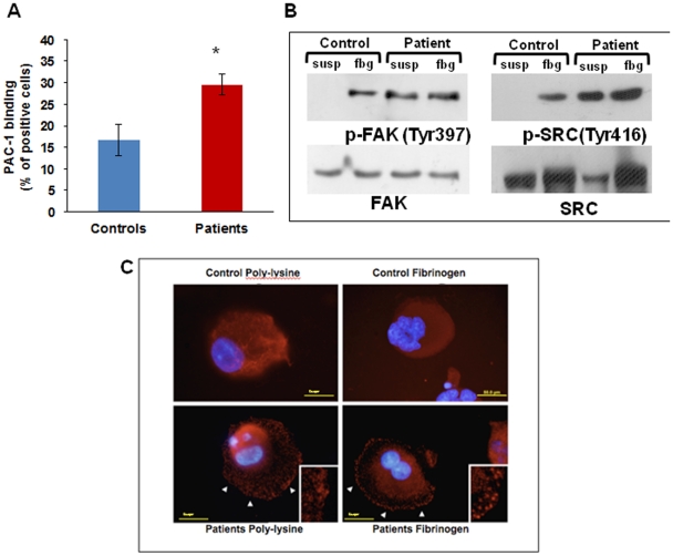
Figure 3. Integrin αIIbβ3 activation and outside-in signalling.
(A) Flow cytometry analysis of PAC-1 binding to resting megakaryocytes is significantly increased in patients as compared with controls. *p<0.05 vs control. (B) Western blotting showed Src and FAK phosphorylation in patient megakaryocytes in suspension as well as after adhesion onto fibrinogen. (C) Differently from control cells (upper panels), patient megakaryocytes showed FAK clustering already after 1 hour of adhesion onto fibrinogen, and also in suspension (lower panels).

