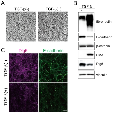
Figure 1. TGF-β-mediated EMT decreases Dlg5 expression.
A: LLc-PK1 cells were incubated with 4 ng/ml of TGF-β for three days. Images were taken using phase contrast microscopy. TGF-β treatment induced morphological changes of LLc-PK1 cells. B: Three days after incubation with TGF-β, cells were lysed and immunoblotted using the indicated antibodies, which include an antibody for the epithelial marker E-cadherin, antibodies for the mesenchymal markers SMA and fibronectin, and antibodies for Dlg5 and β-catenin. As a loading control, vinculin expression was detected. C: LLc-PK1 cells were incubated with 4 ng/ml of TGF-β for three days. The cells were immunostained with anti-Dlg5 or anti-E-cadherin antibody. The scale bar indicates 10 µm. TGF-β treatment induced EMT and decreased Dlg5 expression. The results are representative of at least three independent experiments.

