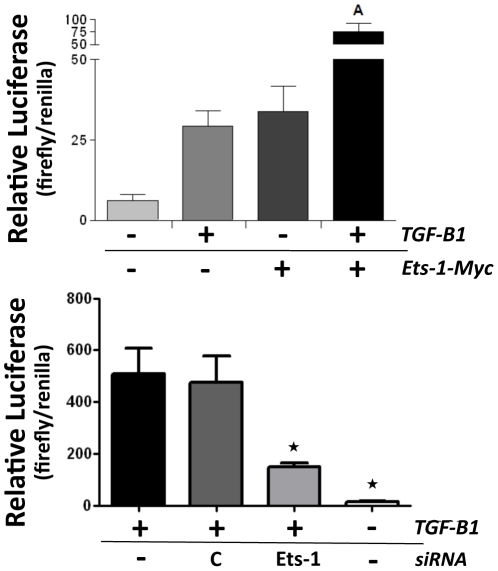
Figure 2. Ets-1 synergizes with TGF-β1 for CCN2 promoter induction in osteoblasts.
(A) Osteoblasts were plated in 96 well tissue culture plates and transfected with either 0.4 µg of an empty vector control (−) or the Ets-1 expression construct (+). All samples were co-transfected with 0.4 µg of our previously described CCN2 promoter luciferase reporter [22] and 0.2 µg of a renilla luciferase expression vector as an internal control. The cells were serum starved for 24 hrs and then treated with TGF-β1 (5 ng/ml) (+) or mock treated (−) with TGF-β1 diluent for 24 hrs. Luciferase activity was then assessed and expressed as a ratio of firefly/renilla luciferase (+SEM, n = 6). A = p<0.05 compared to +TGF-β1 only or +Ets-1 only. (B) Osteoblasts were plated in 96 well tissue culture plates and transfected with either 100 nM of Ets-1 siRNA (Ets-1) or control siRNA (C) for 48 hrs. All samples were co-transfected with 0.4 µg of our previously described CCN2 promoter luciferase reporter [22] and 0.2 µg of a renilla luciferase expression vector as an internal control. The cells were serum starved for 24 hrs and then treated with 5 ng/ml of TGF-β1 (+) or mock treated (−) with TGF-β1 diluent for 24 hrs. Luciferase activity was then assessed and expressed as a ratio of firefly/renilla luciferase (+SEM, n = 6). Star symbol indicates p<0.05 compared to control siRNA.

