Figure 6.

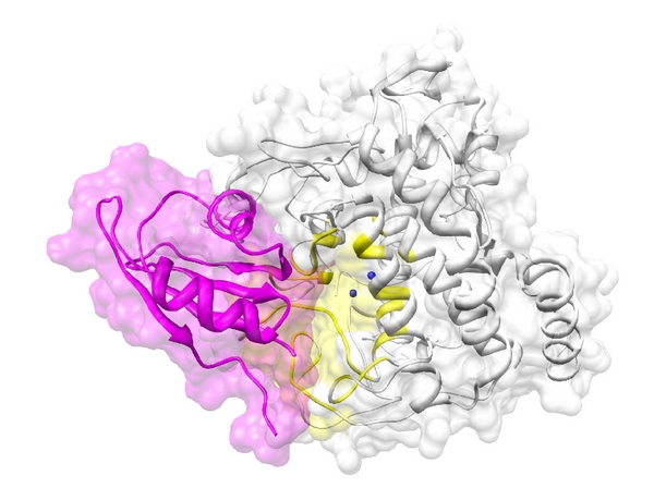
The docking between tyrosinase (white) and RBM9 (magenta). The active site of tyrosinase near two the Cu2+ ions (blue spheres) is colored in yellow. The two proteins are depicted as illustrations.

The docking between tyrosinase (white) and RBM9 (magenta). The active site of tyrosinase near two the Cu2+ ions (blue spheres) is colored in yellow. The two proteins are depicted as illustrations.