
Figure 1.
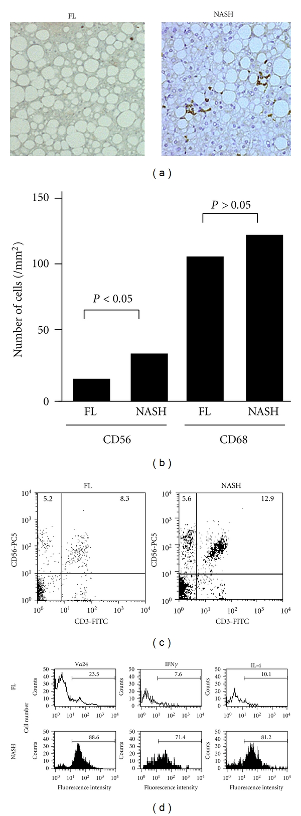
Accumulation of NKT cells in the NAFLD liver with high disease activity. (a) Immunohistochemical study using monoclonal antibody against for CD56 shows accumulation of CD56+ cells in the liver of NAFLD with the disease progresses. (FL; fatty liver versus NASH; nonalcoholic steatohepatitis). (b) The number of CD56+ or CD68+ cells. CD56+ cells are significantly increased as the disease progresses. (c) Flow cytometric analysis of isolated intrahepatic mononuclear cells with NAFLD. Numbers in the quadrant represent the percentage of positive cells. Right-upper quadrant represents NKT cells (CD3+CD56+ cells). (d) Flow cytometric analysis of Vα24 and intracytoplasmic cytokines of gated CD3+CD56+ cells among mononuclear cells isolated from livers with NAFLD. Numbers in each histogram represent the percentage of positive cells. These data have been previously presented in [30].